Blogs
Acoustic Reflex in Otosclerosis: Diagnosis, Causes and Hearing Loss Explained
Acoustic reflex in otosclerosis means the tiny middle ear “protective reflex” that normally tightens the stapes system does not respond as expected because the stapes becomes stiff. In simple terms, the test checks whether the ear’s sound reflex happens and otosclerosis often makes that reflex weak or absent.
This matters because reflex results add an extra clue when someone has gradual hearing loss with otherwise normal looking eardrums. In this article, we explain what acoustic reflex in otosclerosis typically looks like, why it changes, how ENT specialists and audiologists use it in diagnosis and what it can (and cannot) tell you about hearing loss.
Understanding acoustic reflex in otosclerosis
A quick refresher: what happens in otosclerosis?
Otosclerosis is a bone remodelling condition of the otic capsule that commonly affects the stapes footplate. When the stapes becomes fixed, sound vibrations are not transmitted efficiently to the inner ear. This often produces a conductive hearing loss that can slowly worsen over time.
Some people also develop inner ear involvement (cochlear otosclerosis) which can add sensorineural hearing loss and tinnitus. If you want a focused read on that form, see Ascent’s guide on Cochlear Otosclerosis.
What is the acoustic reflex?
The acoustic reflex is an automatic contraction of the stapedius muscle in response to loud sounds. That contraction stiffens the ossicular chain slightly and reduces the amount of sound energy reaching the inner ear, especially for low frequencies.
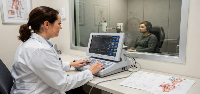
Clinically, the reflex is measured during immittance testing using a probe in the ear canal. This is why acoustic reflex in otosclerosis is discussed alongside tympanometry and middle ear evaluation.
For an overview of the test principles used worldwide in audiology clinics, you can also refer to ASHA’s tympanometry and acoustic reflex guidance.
Why does acoustic reflex in otosclerosis often disappear?
In many patients, acoustic reflex in otosclerosis is absent because the stapes cannot move normally. Even if the stapedius contracts, a fixed stapes limits the measurable change in middle ear compliance.
There is also an “input problem” in conductive hearing loss: less sound energy reaches the cochlea, so the reflex trigger pathway may not receive enough effective stimulation at the levels used for testing.
In practice, clinicians often see absent reflexes with normal ear canal volume and otherwise normal tympanic membrane appearance. That pattern is one reason acoustic reflex in otosclerosis is considered a useful supportive sign, not a stand alone diagnosis.
What does hearing loss feel like in otosclerosis?
People commonly describe:
-
Gradual hearing loss in one ear then both ears
-
Difficulty hearing low pitched voices
-
Tinnitus
-
Better hearing in noisy places (paracusis Willisii) in some patients
These symptoms are not unique to otosclerosis. That is why audiometry, tympanometry and acoustic reflex in otosclerosis are combined with clinical examination by an ENT otologist.
How is acoustic reflex testing performed?
Reflex testing is typically done after tympanometry during an immittance assessment. A probe seals the ear canal, a tone is presented and the instrument measures tiny changes in compliance that indicate a reflex.
Results are usually recorded as:
-
Present at expected levels
-
Present but elevated (needs louder sound)
-
Absent
Because middle ear mechanics strongly influence the measurement, acoustic reflex in otosclerosis is interpreted together with:
-
Pure tone audiometry (air and bone conduction)
-
Tympanometry pattern (Type A, As etc.)
-
Speech testing
-
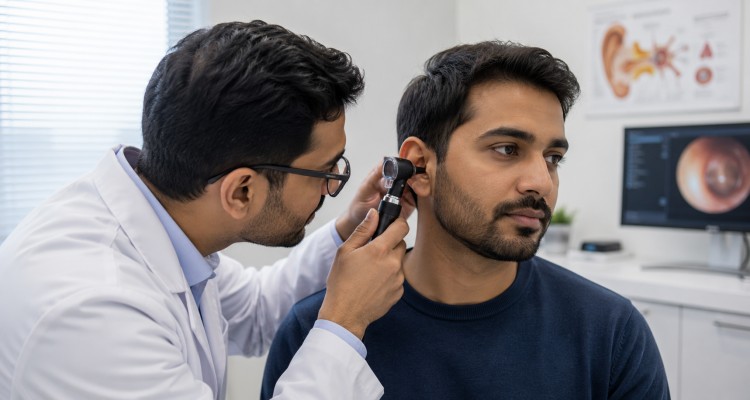
History and otoscopic findings
What patterns do clinicians look for?
A classic teaching point is that stapes fixation tends to reduce compliance and remove reflexes. However, real patients vary. Early otosclerosis can show a near normal tympanogram, so acoustic reflex in otosclerosis can be one of the first “functional” tests that becomes abnormal.
Here is a practical table clinicians use for pattern recognition. These are common patterns, not absolute rules.
| Condition or scenario | Tympanometry (common trend) | Acoustic reflex trend | What it suggests clinically |
|---|---|---|---|
| Normal middle ear | Type A | Present at typical levels | Healthy middle ear function |
| Stapes fixation (otosclerosis) | Type As or Type A | Often absent | Supports acoustic reflex findings in otosclerosis when the audiogram suggests conductive loss |
| Ossicular discontinuity | Type Ad | Often absent or abnormal | Hypercompliant system rather than fixation |
| Middle ear fluid | Type B | Absent | Conductive block due to effusion |
| Large perforation or open tube | Large ECV with flat trace | Not measurable reliably | Consider perforation or ventilation tube |
If an audiogram shows a conductive loss with a Carhart notch and reflexes are absent, acoustic reflex in otosclerosis moves higher on the suspect list. Many ENT teams confirm the diagnosis with a full workup and sometimes CT imaging depending on the case.
Does acoustic reflex in otosclerosis explain the degree of hearing loss?
Acoustic reflex in otosclerosis helps explain the mechanism of hearing loss (stiffened transmission) more than it predicts exact severity. In general, a larger conductive component increases the chance that reflexes will be absent, but the relationship is not perfect.
Also remember that absent reflexes can occur in other conditions, including middle ear effusion and some neural pathway disorders. That is why an ENT otologist focuses on the full clinical picture rather than one test.
How do doctors diagnose otosclerosis step by step?
If you have progressive hearing loss with a normal looking eardrum, a typical evaluation includes:
-
Detailed history (onset, family history, pregnancy related changes, tinnitus)
-
Otoscopy
-
Pure tone audiometry and speech testing
-
Tympanometry plus acoustic reflex in otosclerosis assessment
-
Consideration of imaging (CT temporal bone) when appropriate
A key point is that acoustic reflex in otosclerosis is a supportive test. The diagnosis is usually made by combining audiology patterns with ENT assessment.
What treatments are available when acoustic reflex in otosclerosis is abnormal?
Treatment depends on your hearing levels, lifestyle needs and whether the loss is mainly conductive or mixed.
Hearing aids
Modern digital hearing aids can be very effective for conductive hearing loss, especially when surgery is not preferred or not yet needed.
Stapedotomy or stapedectomy
When stapes fixation is the main cause, stapes surgery can improve sound transmission. If you are exploring this option, you can read about finding a Best Stapedotomy Surgeon in India and what patients typically discuss during surgical counselling.
After successful surgery, the mechanical problem that contributes to acoustic reflex in otosclerosis may change because the ossicular chain mechanics are altered with a prosthesis. Your surgeon and audiologist will explain which tests are useful for follow up in your specific case.
Cochlear implantation in advanced cases
In far advanced otosclerosis with poor aided benefit, cochlear implantation may be considered. This is a specialised decision and should be evaluated by an experienced ENT otologist.
When should you consult an ENT otologist?
Seek an ENT evaluation if you notice:
-
Gradually worsening hearing loss in one or both ears
-
Persistent tinnitus
-
A family history of otosclerosis
-
Difficulty hearing despite normal ear examination elsewhere
At Ascent Hospital patients can access comprehensive ear nose and throat care with advanced diagnostics. Ascent is widely recognised as a best ENT Hospital in Kerala and is Kerala’s first ISO and NABH accredited ENT specialty hospital, with 24/7 ENT emergency support.
If you are looking for an ENT clinic in Kerala or want to consult a Best ENT surgeon in kerala for otosclerosis evaluation, Ascent ENT Hospital Kerala offers end to end assessment including audiology, imaging guidance when needed and surgical expertise.
Conclusion
Acoustic reflex in otosclerosis is a practical clinical clue because otosclerosis stiffens the stapes system and often makes reflexes absent on testing. It does not diagnose otosclerosis alone, but when combined with audiometry and ENT assessment, it helps narrow the cause of hearing loss and guide next steps. Treatment may include hearing aids, stapes surgery or advanced interventions for selected cases.
If you have symptoms that match this pattern, the next best step is a specialist consultation. Book an appointment with Ascent Hospital and schedule your ear evaluation through the contact page to discuss testing, diagnosis and personalised treatment options.
Share
Share on WhatsApp
Our Professionals
 (1) (2).png)
Dr. Sharafudheen. P.K
Chief Consultant ENT & Cochlear Implant Surgeon
MBBS, MS (ENT), DORL DOHNS - RCS Ed (UK)
 (2).jpg)
Dr. Anuradha Varma. M.R
Senior Consultant ENT, Head & Neck Surgeon
MBBS, DLO
.jpeg)
Dr. Bijiraj Vathwam Veettil
Senior Consultant ENT Head & Neck, Sleep Surgeon
MBBS, MS (ENT), Fellowship in Snoring and Sleep apnea Surgeries (Singapore), Fellowship in sinus and skull base surgery
.png)
Dr. Prasanth Parameswaran
Senior Consultant ENT, Head & Neck Surgeon
MBBS, DLO, DNB (ENT), MNAMS, AASC (Specialist - Allergy and Immunotherapy), Fellowship in Snoring and Sleep apnea Surgeries (Singapore).

Dr. Arshad M Razi
Consultant ENT, Head & Neck Surgeon
MBBS, MS (ENT), Fellow in Head & Neck Surgical Oncology

Dr. Deepthi. N.V
Senior Consultant ENT, Head & Neck Surgeon
MBBS, DNB (ENT)


Dr. K Shilpa Nair
ENT Surgeon | Cochlear Implant & Sleep Surgery Specialist
MBBS, MS, DNB, MRCS(Eng), Post doctoral Fellowship in Implantation Otology


Dr. Nibi Shajahan
Consultant ENT, Head & Neck Surgeon
MBBS, MS (ENT), Fellowship in Snoring and Sleep apnea Surgeries (Singapore). Trained in Allergy and Immunotherapy


Dr.Anand Krishnan
Consultant ENT, Head and Neck Surgeon
MBBS ,DLO ,DNB ENT ,MRCS (ENGLAND)


Dr. Seshadri Ganesh. A.L
Senior Consultant Anaesthesiologist
MBBS, DCH, MD, DIP.N.B


Mr. Prasanth. N.P
Senior Audiologist & Speech Language Pathologist
BASLP, MASLP, MSc (Psy)

Ms. Chinju Johny
Audiologist & Speech Language Pathologist
BASLP, MSc (Audiology)


Our Patient Stories
View All Testimonials